Stories|Tracking how Cells absorb Coenzyme Q10
Tracking how Cells absorb Coenzyme Q10
Life Science and healthcare

Tracking How Cells Absorb Coenzyme Q10 – A New View with X-ray Eyes
For the first time, scientists from the University of Hamburg have successfully visualized and quantified how cells take up coenzyme Q10 — a vital molecule for energy production and antioxidant defense — using cutting-edge X-ray fluorescence (XRF) imaging at DESY’s PETRA III synchrotron. This breakthrough not only reveals how Q10 enters cells but also showcases a powerful, non-invasive tool for studying the distribution of other biomolecules and drugs.
Coenzyme Q10, or Ubiquinone, is essential for cellular energy metabolism and protects tissues such as the skin from oxidative stress. Yet, its cellular uptake after supplementation has remained largely mysterious.
Traditional imaging methods like PET or SPECT can track molecules in whole organisms but lack the resolution to see inside individual cells. Other approaches, such as ICP-MS, provide that detail but destroy the sample in the process.
That’s where XRF imaging at PETRA III comes in — offering both high spatial resolution and non-destructive analysis. By using the facility’s intense, precisely tuned X-rays, the researchers achieved a level of sensitivity that finally made it possible to quantify Q10 uptake inside living cells.
To trace Q10’s path, the team created an iodine-labeled version of coenzyme Q10 (I₂-Q10) and introduced it to human skin cells (keratinocytes). After confirming that complementary XRF experiments:
- At beamline P21.1, whole-cell pellets were scanned, revealing that I₂-Q10 accumulated in specific regions within the individual cells — with an uptake signal roughly three times stronger than that of control samples.
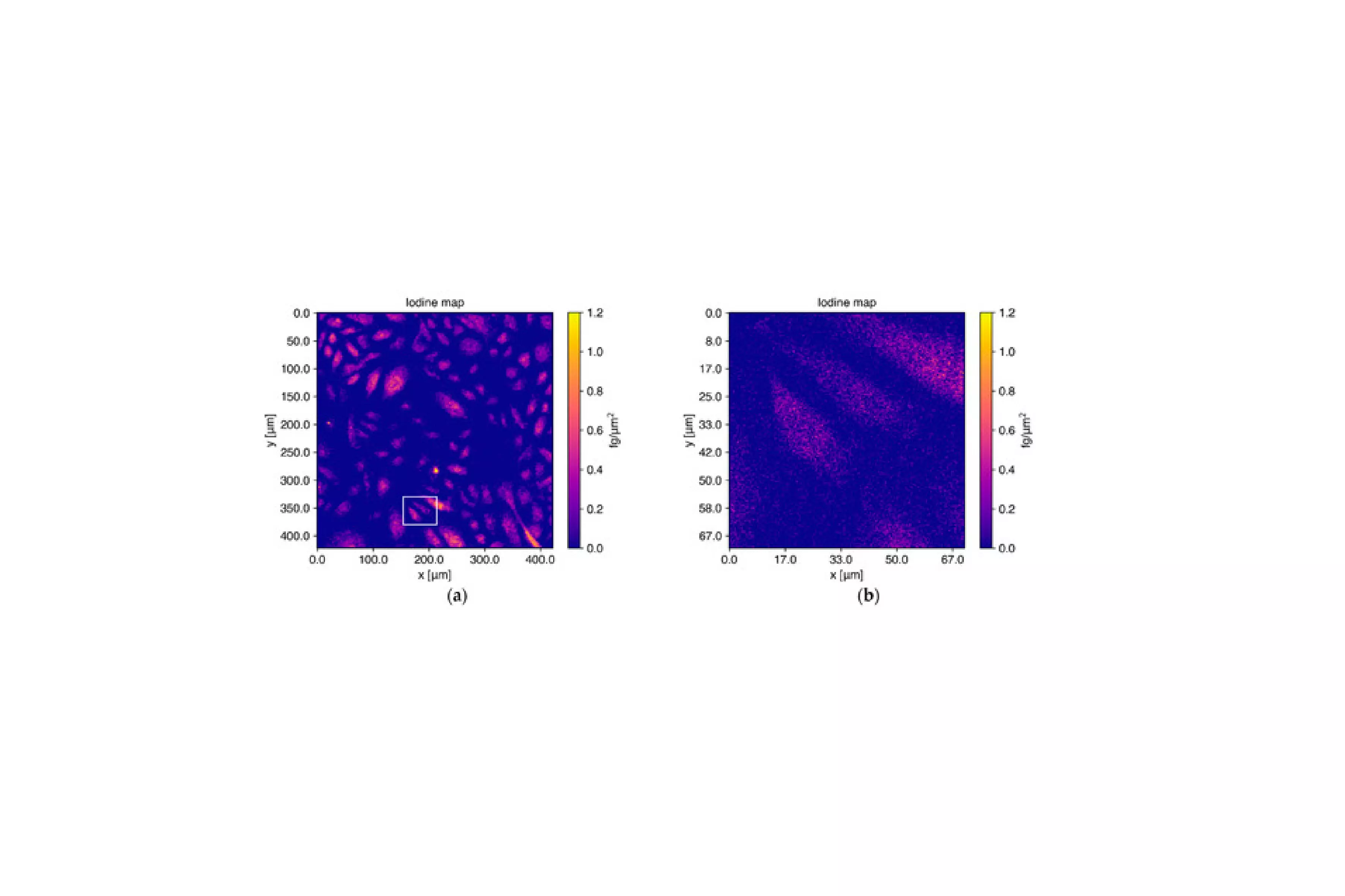
- At beamline P06, sub-cellular mapping was carried out at nanometer resolution. This allowed scientists to pinpoint exactly where Q10 localizes within the cell, switching seamlessly between coarse and fine scans to map hundreds of cells efficiently.
The result: a clear, quantitative picture of Q10’s journey into and through the cell — something no technique had achieved before.
This study demonstrates that synchrotron-based XRF imaging can precisely track how biomolecules are distributed within cells, without destroying them. The approach paves the way for studying drug delivery, bioavailability, and metabolism at an unprecedented level of detail.
With high photon flux and flexible beamline setups, PETRA III offers a unique platform to explore how molecules behave inside living systems — enabling breakthroughs in both biomedical research and pharmaceutical innovation.